-
2021: Reinventing Valentine's Day!

Tired of virtual learning and being locked up for almost a year? Shake things up with these 12 clever ideas!
Posted · Author Arora Periodontics
-
New Year, New Beginnings

victory woman beach Resolutions tend to be easier said than done. So, start by simply trying out something new. Set small milestones, and once you have that habit built up, set a bigger one to accomplish something challenging. Now is the time, after everything we’ve been through this past year, GO FOR IT!
Posted · Author Arora Periodontics
-
There's No Place Like Home For The Holidays
If you’re just not feeling festive, let’s start looking at the holiday season as an exciting challenge, and create new holiday traditions. There are so many fun things to do at home, here are a few of our favorites!

family laying under Christmas tree Posted · Author Arora Periodontics
-
Practicing Gratitude
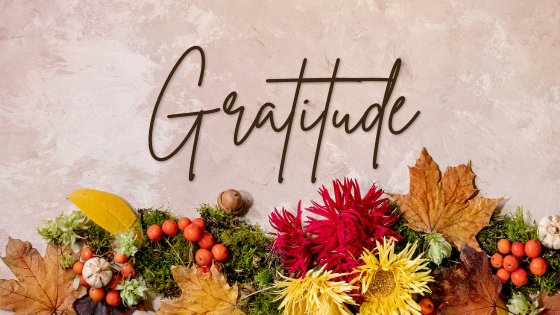
fall bouquet Remember that humans were designed to struggle. So, with confidence and determination, our greatness emerges.
Posted · Author Arora Periodontics
-
Halloween 2020
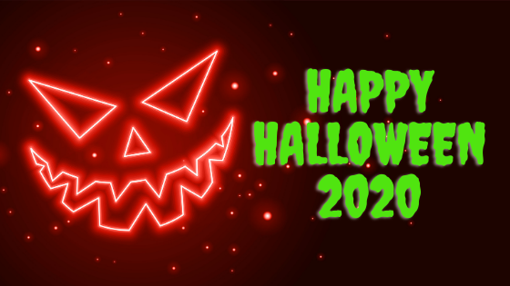
Description = neon jack-o-lantern If you are in a location that has prohibited trick or treating this year, don’t fret! There are plenty of things that you can do during a night in at home...
Posted · Author Arora Periodontics
-
Expectations VS Reality: 4 Dental Myths DEBUNKED!
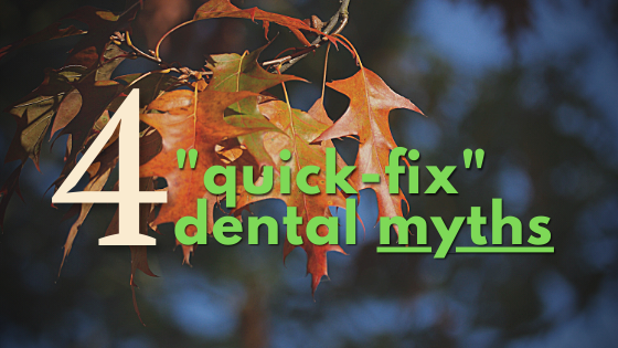
fall leaves on tree Believe it or not, your oral health impacts many different areas of your total well being. Did you know your oral health can contribute to heart disease, diabetes, blood cell disorders, and bacterial pneumonia?
Posted · Author Arora Periodontics